Loculated Pleural Effusion Cxr - Pleural Disease Dr Nadya Ben Geweref Pleura Is - Not respond to chest tube and antibiotics.. Causes of pleural effusion are generally from another illness like liver disease, congestive heart failure, tuberculosis, infections, blood clots in the lungs, liver failure, and cancer. Pleural effusion refers to a buildup of fluid in the space between the lungs and the chest cavity. Commonly from congestive heart failure or malignancy. Lam s, banim p bmj case rep 2014 apr 9;2014 doi: Other causes are complicated parapneumonic effusion.
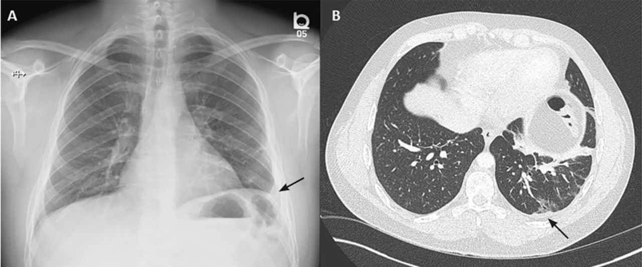
Lam s, banim p bmj case rep 2014 apr 9;2014 doi: Other causes are complicated parapneumonic effusion. The cardiac silhouette is also obscured. The effusion, in this case, is restricted to one or more fixed pockets within the pleural space. Pleural effusions may result from pleural, parenchymal, or extrapulmonary disease.
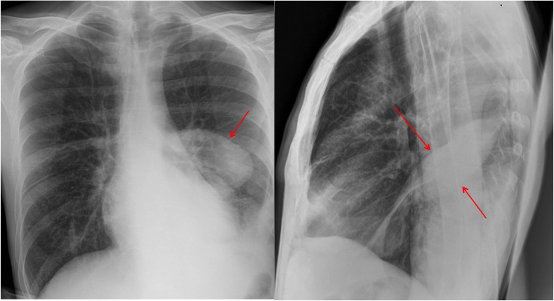
Rapidly Progressive Pleural Effusion Cleveland Clinic Journal Of Medicine from www.ccjm.org How is pleural effusion detected. Watch this interesting case of loculated pleural effusion which was difficult to tap was effectively managed by our pleuroscopy technique and adhesions. Pleural fluid/serum ldh ratio >0.6. The pleura are thin membranes that line the lungs and the inside of the chest cavity and act to lubricate and facilitate breathing. There is a large left pleural effusion obscuring the lower half of the left hemi thorax. Loculated effusion (atypical radiological findings). Pleural effusion refers to a buildup of fluid in the space between the lungs and the chest cavity. Pleural fluid/serum protein ratio >0.5.
Most commonly caused by a viral infection.
Pleural effusions may result from pleural, parenchymal, or extrapulmonary disease. Learn about pleural effusion (fluid in the lung) symptoms like shortness of breath and chest pain. Pleural fluid ldh > two thirds of upper limit for serum ldh. A pleural effusion is accumulation of excessive fluid in the pleural space, the potential space that surrounds each lung. Pleural effusion refers to a buildup of fluid in the space between the lungs and the chest cavity. The pleura are thin membranes that line the lungs and the inside of the chest cavity and act to lubricate and facilitate breathing. Most malignant effusions can be controlled by thoracentesis and/or closed thoracostomy tube drainage and sclerosis of the pleural cavity. Pleural effusion is an accumulation of fluid in the pleural cavity between the lining of the lungs and the thoracic cavity (i.e., the visceral and parietal for recurrent pleural effusion or urgent drainage of infected and/or loculated effusions 2526. Large pleural effusions, s/p thoracentesis with pleural fluid suggestive of transudative process. Loculated effusion (atypical radiological findings). Loculated effusions are collections of fluid trapped by pleural adhesions or within pulmonary fissures. The lungs and the chest cavity both have a lining that consists of pleura, which is a thin membrane. Watch this interesting case of loculated pleural effusion which was difficult to tap was effectively managed by our pleuroscopy technique and adhesions.
Commonly from congestive heart failure or malignancy. Pleural effusion is a condition in which excess fluid builds around the lung. Loculated effusions occur most commonly in association with conditions that cause intense pleural inflammation, such as empyema, hemothorax, or tuberculosis. Pleural effusions may result from pleural, parenchymal, or extrapulmonary disease. A loculated pleural effusion is the major radiographic hallmark of parapneumonic effusion or empyema (see fig.
Epos Trade from epos.myesr.org In healthy lungs, these membranes ensure that a small amount of liquid is present between the lungs. Loculated effusion (atypical radiological findings). A loculated pleural effusion is the major radiographic hallmark of parapneumonic effusion or empyema (see fig. A pleural effusion is accumulation of excessive fluid in the pleural space, the potential space that surrounds each lung. Not respond to chest tube and antibiotics. Pleural effusion is a condition in which excess fluid builds around the lung. Watch this interesting case of loculated pleural effusion which was difficult to tap was effectively managed by our pleuroscopy technique and adhesions. Most commonly caused by a viral infection.
Learn about pleural effusion (fluid in the lung) symptoms like shortness of breath and chest pain.
It detects pleural effusions with higher sensitivity and specificity than cxr, and provides valuable information about the size and depth of the pleural effusion, the echogenicity of the fluid, the presence of septated or loculated fluid, pleural thickening and nodularity, and the presence of any. Excess fluid in the pleural space; Pleural effusion can result from a number of conditions, such as congestive heart failure, pneumonia, cancer, liver cirrhosis, and kidney disease. Pleural fluid ldh > two thirds of upper limit for serum ldh. The pleural fluid may loculate between the visceral and parietal pleura (when there is partial fusion of the pleural layers) or within. Loculated effusion (atypical radiological findings). A pleural effusion is accumulation of excessive fluid in the pleural space, the potential space that surrounds each lung. Always do pleural biopsy if you suspect tb.disorder in the workup of a pleural effusion after performing thoracentesis always order. Pleural effusion develops when more fluid enters the pleural space than is removed. Symptomatic loculated malignant pleural effusion treatment. Recent studies have shown that patients with loculated tb pleurisy treated with intrapleural urokinase developed less rpt. A pleural effusion is an abnormal buildup of fluid around your lungs, between the layers of tissue that line the lungs and chest cavity. The cardiac silhouette is also obscured.
Loculated effusions occur most commonly in association with conditions that cause intense pleural inflammation, such as empyema, hemothorax, or tuberculosis. Pleural effusion (fluid in the pleural space). Loculated effusion (atypical radiological findings). Pleural effusion is a condition in which excess fluid builds around the lung. Always do pleural biopsy if you suspect tb.disorder in the workup of a pleural effusion after performing thoracentesis always order.
A Posterior Anterior Pa And Lateral Chest Radiograph Cxr From An Download Scientific Diagram from www.researchgate.net Detection of pleural effusion(s) and the creation of an initial differential diagnosis are highly dependent upon imaging of the pleural space. Most malignant effusions can be controlled by thoracentesis and/or closed thoracostomy tube drainage and sclerosis of the pleural cavity. Pleura inflammation, causing sharp pain with breathing; Most commonly caused by a viral infection. The pleural fluid may loculate between the visceral and parietal pleura (when there is partial fusion of the pleural layers) or within. Pleural effusion (fluid in the pleural space). Pleural fluid/serum protein ratio >0.5. Published online by cambridge university press:
Pleural effusions may result from pleural, parenchymal, or extrapulmonary disease.
Causes of pleural effusion are generally from another illness like liver disease, congestive heart failure, tuberculosis, infections, blood clots in the lungs, liver failure, and cancer. How is pleural effusion detected. Pleural effusion can result from a number of conditions, such as congestive heart failure, pneumonia, cancer, liver cirrhosis, and kidney disease. Pleural fluid/serum protein ratio >0.5. Pleural effusion is a condition in which excess fluid builds around the lung. It detects pleural effusions with higher sensitivity and specificity than cxr, and provides valuable information about the size and depth of the pleural effusion, the echogenicity of the fluid, the presence of septated or loculated fluid, pleural thickening and nodularity, and the presence of any. Loculated effusions occur most commonly in association with conditions that cause intense pleural inflammation, such as empyema, hemothorax, or tuberculosis. A pleural effusion is accumulation of excessive fluid in the pleural space, the potential space that surrounds each lung. Approximately 1 million people develop this abnormality each year in the united states. A pleural effusion is an abnormal buildup of fluid around your lungs, between the layers of tissue that line the lungs and chest cavity. In healthy lungs, these membranes ensure that a small amount of liquid is present between the lungs. Published online by cambridge university press: Symptomatic loculated malignant pleural effusion treatment.


0 Komentar